Abdomen Anatomy-Female : Animation Anatomy Of Female Abdomen - The abdominal cavity is the part of the body that houses the stomach, liver, pancreas, kidneys, gallbladder, spleen, and the large and small intestines.
Abdomen Anatomy-Female : Animation Anatomy Of Female Abdomen - The abdominal cavity is the part of the body that houses the stomach, liver, pancreas, kidneys, gallbladder, spleen, and the large and small intestines.. The diaphragm marks the top of the abdomen and the horizontal line at the level of the top of the pelvis marks the bottom. The true pelvis, or lesser pelvis, lies below the pelvic brim (figure 1). One major difference between males and females. Anterolateral and posterior abdominal walls. The region of the trunk that lies between the diaphragm above the pelvic inlet below is referred to as abdomen that is divided into nine regions by two vertical and two horizontal lines.
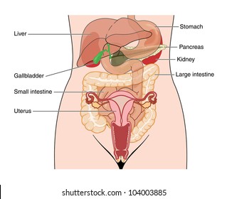
It is a highly muscular, childbearing organ in females, approximating 3 x 2 x 1 inches in size in a nulliparous. These two apertures, together with abdominal walls, bound the abdominal cavity. This medical exhibit diagram illustrates the anatomy of the female abdomen and pelvis from an anterior front cut away view showing elements of the digestive system. Those organs include the stomach, small intestine, colon, liver, gallbladder, spleen, and pancreas. If we keep in mind some common issues like, the pain of acidity is in the upper part of the abdomen.
Abdomen And Pelvis 3d Interactive Anatomy Tutorials from anatomyzone.com The muscles of the abdomen protect vital organs underneath and provide structure for the spine. The abdomen is the body region found between the thorax and the pelvis. The abdominal wall surrounds the abdominal cavity, providing it with flexible coverage and protecting the internal organs from damage. These structures collectively fall under the term vulva. Summary female anatomy includes the external genitals, or the vulva, and the internal reproductive organs, which include the ovaries and the uterus. The majority of these organs. Abdominal computed tomography (ct) is a type of medical imaging procedure used to diagnose and monitor internal stomach issues, like cancer, bowel obstruction, and abdominal pain. The major organs of the abdomen include the.
The true pelvis, or lesser pelvis, lies below the pelvic brim (figure 1).
Those organs include the stomach, small intestine, colon, liver, gallbladder, spleen, and pancreas. The muscles of the abdomen protect vital organs underneath and provide structure for the spine. At the level of the pelvic bones, the abdomen. Über 7 millionen englischsprachige bücher. The region of the trunk that lies between the diaphragm above the pelvic inlet below is referred to as abdomen that is divided into nine regions by two vertical and two horizontal lines. The abdominal cavity is the part of the body that houses the stomach, liver, pancreas, kidneys, gallbladder, spleen, and the large and small intestines. Anatomy, abdomen and pelvis, female pelvic cavity; The major muscles of the abdomen include the rectus. This medical exhibit diagram illustrates the anatomy of the female abdomen and pelvis from an anterior front cut away view showing elements of the digestive system. Those structures are contained in its cavity, the abdominal cavity. If we keep in mind some common issues like, the pain of acidity is in the upper part of the abdomen. The muscles of the lower back, including the erector spinae and quadratus lumborum muscles, contract to extend and laterally bend the vertebral column. The diaphragm forms the upper surface of the abdomen.
The abdomen (commonly called the belly) is the body space between the thorax (chest) and pelvis. It is a flexible dynamic container, housing most of the organs of the digestive system and part of the urogenital system. The abdominal muscles also play a major role in the posture and stability to the body and compress the organs of the abdominal cavity during various activities such as breathing and defecation. The liver stomach and abdominal contents are clearly identified and labeled including the cecum ascending colon transverse colon descending colon and small. Related posts of abdominal anatomy of female abdominal regions and organs.
Mobile Site Preview Human Body Organs Abdominal Muscles Anatomy Body Organs Diagram from i.pinimg.com The abdominal wall surrounds the abdominal cavity, providing it with flexible coverage and protecting the internal organs from damage. Anatomy, abdomen and pelvis, female pelvic cavity; Those structures are contained in its cavity, the abdominal cavity. The liver stomach and abdominal contents are clearly identified and labeled including the cecum ascending colon transverse colon descending colon and small. Those organs include the stomach, small intestine, colon, liver, gallbladder, spleen, and pancreas. At the level of the pelvic bones, the abdomen. If you plan to enter a healthcare profession such as nursing, this is something you'll use on the job when performing abdominal assessments (and while documenting). It is bounded superiorly by the xiphoid process and costal margins, posteriorly by the vertebral column and inferiorly by the pelvic bones and inguinal ligament.
Anterolateral and posterior abdominal walls.
These muscles help the body bend at the waist. One major difference between males and females. The liver stomach and abdominal contents are clearly identified and labeled including the cecum ascending colon transverse colon descending colon and small. Gallbladder pain is below the ribs on the upper right side of the abdomen. Über 7 millionen englischsprachige bücher. The abdomen is comprised primarily of the digestive tract and other accessory organs which assist in digestion, the urinary system, spleen, and the abdominal muscles (shown below). The abdomen is the region of the trunk between the thorax and the pelvis. This landmark begins at the level of the sacral promontory posteriorly. The abdominal wall can be divided into two sections: The major muscles of the abdomen include the rectus abdominis in front the external obliques at the sides and the latissimus dorsi muscles in the back. The region of the trunk that lies between the diaphragm above the pelvic inlet below is referred to as abdomen that is divided into nine regions by two vertical and two horizontal lines. Inferiorly the abdomen is open to the pelvis, communicating through the superior pelvic aperture (pelvic inlet). In women, the lowest portion of the abdomen is actually the pelvis and involves the uterus,.
The region of the trunk that lies between the diaphragm above the pelvic inlet below is referred to as abdomen that is divided into nine regions by two vertical and two horizontal lines. The diaphragm forms the upper surface of the abdomen. One major difference between males and females. Anatomy, abdomen and pelvis, female pelvic cavity; Those organs include the stomach, small intestine, colon, liver, gallbladder, spleen, and pancreas.
Abdominal Organs Images Stock Photos Vectors Shutterstock from image.shutterstock.com The female reproductive system is an intricate arrangement of structures that can separate into external and internal genitalia. The diaphragm marks the top of the abdomen and the horizontal line at the level of the top of the pelvis marks the bottom. The majority of these organs. Four abdominal quadrants and nine abdominal regions in anatomy and physiology, you'll learn how to divide the abdomen into nine different regions and four different quadrants. This medical exhibit diagram illustrates the anatomy of the female abdomen and pelvis from an anterior front cut away view showing elements of the digestive system. The abdomen is the body region found between the thorax and the pelvis. Anterolateral and posterior abdominal walls. Anatomy, abdomen and pelvis, female pelvic cavity;
Those organs include the stomach, small intestine, colon, liver, gallbladder, spleen, and pancreas.
The abdominal wall can be divided into two sections: The female reproductive system is an intricate arrangement of structures that can separate into external and internal genitalia. The diaphragm forms the upper surface of the abdomen. The abdomen is comprised primarily of the digestive tract and other accessory organs which assist in digestion, the urinary system, spleen, and the abdominal muscles (shown below). If we keep in mind some common issues like, the pain of acidity is in the upper part of the abdomen. This medical exhibit diagram illustrates the anatomy of the female abdomen and pelvis from an anterior front cut away view showing elements of the digestive system. Anterolateral and posterior abdominal walls. The muscles of the abdomen protect vital organs underneath and provide structure for the spine. Summary female anatomy includes the external genitals, or the vulva, and the internal reproductive organs, which include the ovaries and the uterus. The abdominal muscles also play a major role in the posture and stability to the body and compress the organs of the abdominal cavity during various activities such as breathing and defecation. These two apertures, together with abdominal walls, bound the abdominal cavity. It is a highly muscular, childbearing organ in females, approximating 3 x 2 x 1 inches in size in a nulliparous. The diaphragm marks the top of the abdomen and the horizontal line at the level of the top of the pelvis marks the bottom.


0 Komentar